
Abstract
We present the case of a 63-year-old female who developed delayed cognitive and behavioural deterioration secondary to suspected toxic-hypoxic leukoencephalopathy following an intentional oxycodone overdose. Initial neuroimaging was nonspecific, but subsequent evaluations revealed a subacute infarct and diffuse white matter changes. Comprehensive investigations excluded other potential causes, and her condition gradually improved with supportive management and neuro rehabilitation. This case underscores the need to consider delayed toxic hypoxic leukoencephalopathy as a differential diagnosis in patients who develop progressive neuropsychiatric symptoms following an opioid overdose.
Keywords
Drug Overdose, Hypoxia, Leukoencephalopathy, Neuropsychiatry, Oxycodone, White Matter
Introduction
Delayed toxic-hypoxic leukoencephalopathy (DTHL) is a rare neurological syndrome characterized by delayed neuropsychiatric deterioration following an initial recovery from a hypoxic or toxic event. Common triggers include opioid overdose, carbon monoxide exposure, and other neurotoxic substances.1,2 The condition is typically characterized by a biphasic clinical course: an initial recovery phase followed by a subacute onset of cognitive impairment, behavioural changes, and motor dysfunction, which occurs days to weeks later.3 Magnetic resonance imaging (MRI) typically reveals widespread white matter hyperintensities, with sparing of the cortex, in the early stages, and is the key diagnostic tool for identifying the condition. The pathophysiology is believed to involve delayed demyelination resulting from oligodendrocyte damage and secondary disruption of the blood-brain barrier.4,5 Diagnosis is often challenging due to the nonspecific symptoms and the delayed nature of presentation. A thorough exclusion of mimics such as stroke, autoimmune encephalitis, paraneoplastic syndromes, and hereditary leukodystrophies is necessary.6 The prognosis of delayed toxic hypoxic leukoencephalopathy (DTHL) varies, with some patients experiencing full recovery while others may have persistent neurological deficits or even death.7
This report describes a case of a 63-year-old female who developed delayed neurocognitive decline and behavioural disturbances nearly four weeks after an intentional oxycodone overdose. It highlights the clinical course, diagnostic process, and recovery support necessary in the treatment of a patient with medical and psychiatric comorbidities and diagnosis of DTHL.
Case Presentation
A 63-year-old Caucasian female with a history of depression and anxiety disorder, known to the adult community mental health team and diagnosed with Hashimoto’s thyroiditis and diverticular disease was brought to the emergency department by paramedics following a suspected intentional overdose of an unknown medication in an undetermined quantity. On initial assessment by paramedics, she presented with a Glasgow Coma Scale (GCS) score of 4 (E1V2M1), non-reactive pinpoint pupils, and shallow respirations (respiratory rate: 8 breaths per minute). She was bradycardic (heart rate: 45 bpm), hypotensive (BP: 78/52 mmHg), hypoxic (SpO₂: 79%), and hypothermic (temperature: 34.8°C). During pre-hospital care, she received intravenous fluids, high-flow oxygen at 15 L/min, and three doses of naloxone (400 mcg each).
Upon arrival to the emergency department (ED), her GCS had improved to 8, and she was able to maintain her airway; however, she remained bradycardic (heart rate: 37 bpm), hypoxic (SpO₂: 90% on 2 L O₂), and hypotensive (SBP: 70 mmHg), necessitating intravenous fluids, atropine, and a metaraminol infusion.
 During further evaluation in the ED, she was noted to have right- sided hemiparesis, prompting urgent Computed Tomography (CT) brain and CT angiogram of the Circle of Willis and neck vessels and review by the stroke team. Neuroimaging revealed no evidence of an intracranial haemorrhage or central thrombotic arterial occlusion of the major branches of the Circle of Willis. The intensive care team was consulted and recommended a trial of naloxone infusion. Following administration, her GCS improved to 14, and upon regaining full consciousness, she disclosed ingesting an unknown quantity of oxycodone with suicidal intent. A continuous naloxone infusion was initiated at 400 mcg/hour.
 The patient was enrolled in the medical ward for further inpatient management and monitoring of their condition. During her inpatient stay, she was referred to the Consultation-Liaison Psychiatry team for mental health evaluation. Once medically stabilized, she was transferred to the mental health unit for further psychiatric assessment and care.
She initially settled well into the ward environment and was regularly reviewed by the treating psychiatrist. During these reviews, she reported escalating anxiety symptoms, which were attributed to multiple ongoing psychosocial stressors. Her psychotropic regimen was subsequently optimized with sertraline, mirtazapine, and initiating risperidone. She also received input from clinical psychology and occupational therapy teams during her inpatient stay. In the early phase of admission, she was oriented, displayed mostly a euthymic affect but occasionally anxious, actively engaged with peers, and participated fully in structured ward-based activities such as art, yoga, and hydrotherapy. She remained independent in all activities of daily living.
However, on day 26 following the overdose, she experienced an acute behavioural shift marked by cognitive and functional decline. She became vague and confused, with fluctuating awareness of time, place, and person. She was noted to be inattentive and apathetic along with diminished spontaneous movements, limited verbal responsiveness, and significantly reduced interaction with staff, peers, and family members. She started requiring full assistance with activities of daily living, including showering and dressing.
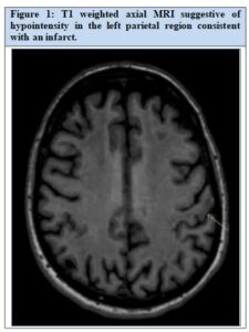
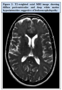
The initial brain MRI revealed symmetric supratentorial parenchymal signal changes, which were nonspecific and indeterminate for leukoencephalopathy. A neurology consultation was obtained, and a clinical examination revealed no focal neurological deficits. A repeat contrast-enhanced MRI of the brain demonstrated cortical and gyri form enhancement in the left postcentral gyrus, consistent with a subacute infarct and a breakdown in the blood-brain barrier (Fig 1 and 2). A late subacute infarct was shown in the left temporal lobe. There was no change in the asymmetric diffuse hyperintensities in the cerebral white matter, suggestive of leukoencephalopathy. The patient’s clinical presentation and neuroimaging findings were reviewed at the multidisciplinary team meeting in neuroradiology. It was concluded that the observed changes in imaging were consistent with a maturing infarct. A retrospective review of the CT brain angiogram obtained at the time of admission revealed features suggestive of a likely left parietal stroke. The infarct was considered the likely cause of the patient’s acute neurological deterioration with delayed onset following the opioid overdose. At the time, the aetiology of the diffuse white matter hyperintensities remained unclear.
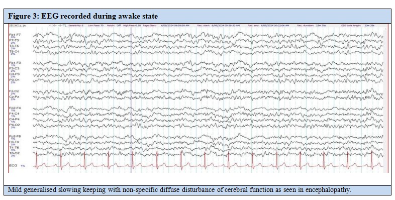
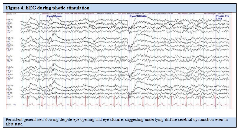
Electroencephalography (EEG) showed mild generalized slowing, indicative of a nonspecific diffuse disturbance in cerebral function consistent with encephalopathy, with no evidence of epileptiform activity (Fig 3 and 4).
Over the course of her inpatient stay, the patient developed progressive gait instability, impaired coordination, and urinary incontinence. Her apathy and psychomotor slowing advanced to a state of abulia. Meanwhile, her mental state remained stable, with no evidence of depressive symptoms or anxiety.
Comprehensive blood investigations were unremarkable. Toxicology screening was negative. Autoimmune markers, including rheumatoid factor, C3/C4 complement levels, cryoglobulins, erythrocyte sedimentation rate (ESR), rapid plasma reagin (RPR), and antinuclear antibodies (ANA), were all within normal limits (Table 1). Cerebrospinal fluid (CSF) analysis revealed normal protein levels and leukocyte counts. CSF cytology, as well as testing for serum and CSF oligoclonal bands, N-methyl-D- aspartate (NMDA) receptor antibodies, voltage-gated potassium channel (VGKC) antibodies, and alpha-amino-3-hydroxy-5-methyl-4-isoxazolepropionic acid (AMPA) receptor antibodies, returned negative results. Paraneoplastic antibody screening in serum was also negative. Genetic testing for leukodystrophies revealed no abnormalities. Finally, a CT scan of the chest, abdomen, and pelvis excluded the presence of any occult malignancy.
 Around three weeks following the onset of behavioural and neurological decline, the patient began to exhibit gradual improvement in cognitive function and engagement. A repeat brain MRI showed no significant change in the leukoencephalopathy and persistent findings related to the prior infarcts. To further evaluate the cerebral white matter abnormalities, an MRI of the spinal cord was done and reported as normal. A follow-up EEG also demonstrated normal findings.
 A neuropsychological assessment was conducted. Mood symptoms were screened using the 21-item Depression, Anxiety, and Stress Scale (DASS-21),8 which yielded results within normal limits. To assess behavioural changes associated with potential frontal lobe dysfunction, the Frontal Systems Behaviour Scale (FrSBe)9 was administered. The patient demonstrated significant difficulty completing the self-report component, and ratings provided by a collateral informant revealed clinically significant abnormalities across all three domains: apathy, disinhibition, and executive dysfunction. Cognitive testing indicated performance below premorbid expectations across multiple domains. Specific deficits—classified as ‘below average’ or lower—were observed in attention, learning, memory, working memory, processing speed, executive function, and visuospatial construction. These impairments were found to interfere with instrumental activities of daily living. The overall profile was consistent with a diagnosis of
mild neurocognitive disorder due to multiple aetiologies with behavioural disturbance, by criteria outlined in the Diagnostic and Statistical Manual of Mental Disorders, Fifth Edition (DSM-5).10
Following comprehensive neurological evaluations and diagnostic investigations, subacute leukoencephalopathy, likely delayed toxic hypoxic leukoencephalopathy, was identified as the most likely cause of the patient’s acute cognitive and behavioural deterioration. The neurology team initiated aspirin 100 mg once daily. Supportive care measures were also provided, including physiotherapy, adequate hydration, and neurocognitive monitoring. Corticosteroids and immunomodulatory therapies were not administered.
The patient demonstrated gradual clinical improvement, with resolution of motor symptoms and regained orientation. She was able to resume performing activities of daily living independently. While cognitive function did not return to premorbid levels, partial improvement was noted. As there were no clinical signs suggestive of anxiety or depression, psychotropic medications were tapered accordingly.
A multidisciplinary discharge planning meeting was conducted involving the treating team, neurology, allied health professionals, and the patient’s family. It was recommended that she undergo a neurology outpatient follow-up in six months, with a repeat brain MRI to assess progress. Post-discharge, she was referred to the adult community mental health team for continued monitoring of her mental state. She was also enrolled in the Rehab in the Home (RITH) program, with occupational therapy sessions scheduled twice weekly for 12–16 weeks to support ongoing cognitive rehabilitation in her home environment. A referral was initiated to Neurocare to offer carer support and promote engagement with outpatient services. A plan was established to closely monitor her cognitive function and explore an application to the National Disability Insurance Scheme (NDIS)11 for further support services.
Discussion
This case demonstrates the classic biphasic course of DTHL, beginning with a life-threatening opioid overdose followed by a delayed onset of cognitive and behavioural decline. The patient’s initial improvement post-overdose and subsequent abrupt deterioration on day 26 are characteristic of this syndrome.2,3
MRI findings in this case, namely, diffuse white matter hyperintensities and delayed cortical/gyriform enhancement are consistent with reported imaging patterns in DTHL and suggest a combined effect of hypoxic injury and infarction.4,12 Although her early brain imaging was inconclusive, repeat imaging combined with clinical deterioration and EEG changes ultimately supported the diagnosis. Retrospective CT angiogram review also indicated a likely infarct, adding complexity to the clinical picture.
Neuropsychological assessment revealed impairments in multiple cognitive domains, including executive function, memory, attention, and visuospatial skills, – findings which are frequently seen in toxic or hypoxic leukoencephalopathy.13 The use of the Frontal Systems Behaviour Scale (FrSBe),9 and DASS-21,8 helped to characterize her behavioural profile and distinguish neuropsychiatric features from mood symptoms. Notably, her cognitive profile met DSM-5 criteria for mild neurocognitive disorder due to multiple aetiologies.10
The management of DTHL remains largely supportive. Corticosteroids and immunotherapy have been explored in isolated cases but with limited supporting evidence.14 In this patient, the use of aspirin, neurorehabilitation, and structured follow-up allowed for partial recovery of motor function and improvement in orientation and daily functioning. However, persistent cognitive deficits highlight the often incomplete recovery associated with DTHL, particularly in older patients.15 The prognosis of DTHL is highly variable and depends on several factors including duration of hypoxic insult, extent of white matter involvement, and timeliness of supportive care. While some patients recover fully over weeks to months, others experience persistent neurocognitive deficits, psychiatric symptoms, or motor impairments.16
This case emphasizes the significance of a multidisciplinary approach that includes neurology, psychiatry, allied health professionals, and family support. A key aspect of her ongoing care involved integrating cognitive rehabilitation with discharge planning, which included utilizing community mental health services and occupational therapy.
Conclusion
The DTHL is an infrequent but clinically important neurological condition that warrants consideration in patients presenting with subacute cognitive and behavioural deterioration following a temporary recovery from hypoxic or toxic insult. Timely diagnosis supported by neuroimaging, systematic exclusion of differential diagnoses, and integrated multidisciplinary care are key to improving clinical outcomes. Although full neurological recovery may not always be attainable, early targeted rehabilitation and engagement with community services can lead to meaningful enhancements in functional capacity and quality of life.
Ethical considerations
Written informed consent was obtained from the patient for the publication of this case report and any associated images.
Acknowledgement
The authors would like to thank the medical team, allied health team and community mental health team involved in the care of the patient. We are also thankful to the patient who consented to share their case for educational and academic purposes.
Author information: Ann Maria Varghese, MBBS, MHS (Neuroscience), Psychiatry Registrar, St John of God Midland Public and Private Hospital, 1 Clayton Street, Midland, Western Australia 6056, Email: [email protected], ORCID:0009-0000-1525-2380; Stephanus Schutte, MBChB, FRANZCP, Consultant Psychiatrist, Adjunct position: Senior Lecturer University of Western Australia, St John of God Midland Public and Private Hospital, 1 Clayton Street, Midland, Western Australia 6056, Email: [email protected], ORCID: 0009-0009-0602-3426; Jayashree Viswanathan, MBBS, MD, MRCPsych, MSc, FRANZCP, Consultant Psychiatrist, St John of God Midland Public and Private Hospital, 1 Clayton Street, Midland, Western Australia 6056, Email: [email protected] ORCID: 0009-0003-7593-3753 |
Correspondence: Jayashree Viswanathan, St John of God Midland Public and Private Hospital, 1 Clayton Street, Midland, Western Australia 6056. Email: [email protected] |
Competing interests: None |
Funding: None |
Received:: 17 June 2025; Revised: 8 July 2025; Accepted: 16 July 2025; Published: 18 July 2025 |
Copyright: © 2025 The Author(s). This is an open-access article distributed under the terms [CC BY-NC] which permits unrestricted use, distribution, and reproduction in any medium, provided the original author and source are credited. |
Citation: Varghese AM, Schutte S, Viswanathan J. Delayed toxic-hypoxic leukoencephalopathy following oxycodone overdose: a case report. Journal of Geriatric Care and Research, 2025, 12, 2: 37-42. |
References
- Shprecher DR, Mehta L. The syndrome of delayed post-hypoxic leukoencephalopathy. NeuroRehabilitation. 2010;26(1):65–72.
- Wijdicks EF. Delayed post-hypoxic leukoencephalopathy: a syndrome of diffuse white matter injury. Neurology. 2009;72(11):900–1.
- Filley CM, Kleinschmidt-DeMasters BK. Toxic leukoencephalopathy. N Engl J Med. 2001;345(6):425–32.
- Lee HB, Yoon PH, Kim DI, et al. Delayed post-hypoxic leukoencephalopathy: MR imaging findings. Korean J 2001;2(2):114–7.
- BanoS, Chaudhary V, Garga Imaging findings in delayed posthypoxic leukoencephalopathy: A case report and review of literature. Indian J Radiol Imaging. 2014;24(4):354–7.
- Finkelstein Y, Aks SE, Hutson JR, Juurlink DN. Opioidtoxicity: Clinical management. In: Brent J, Wallace KL, Burkhart KK, et al., editors. Critical Care Toxicology. 2nd ed. New York: Springer; 2017. 1117–32.
- Beeskow D, Oberstadt M, Saur D, Hoffmann KT, Lobsien D. Delayed post-hypoxic leukoencephalopathy (DPHL)—An uncommon variant of hypoxic brain injury. Front Neurol [Internet].2018 [cited 2025 Jun 10];9:708. Available from: https://www.frontiersin.org/articles/10.3389/fneur.2 00708/full
- Lovibond SH, Lovibond PF. Manual for the Depression Anxiety Stress Scales. 2nd ed. Sydney: Psychology Foundation; 1995.
- Grace J, Malloy PF. Frontal Systems Behavior Scale (FrSBe): Professional manual. Lutz (FL): Psychological Assessment Resources; 2001.
- American Psychiatric Association. Diagnostic and statistical manual of mental disorders. 5th ed. Arlington (VA): American Psychiatric Publishing; 2013.
- National Disability Insurance Scheme. National Disability Insurance Scheme [Internet]. Canberra: National Disability InsuranceAgency; [cited 2025 Jun 10]. Available from: https://www.ndis.gov.au/
- Espinosa PS, Sobrinho M, Martinez R. Delayed toxic leukoencephalopathy after heroin overdose. Clin Neurol Neurosurg. 2015;130:103–6.
- Cummings JL. Frontal-subcortical circuits and human behavior. Arch Neurol. 1993;50(8):873–80.
- Scullen T, Mathkour M, Carr C, Keen J, Dallapiazza RF. Delayed leukoencephalopathy after heroin intoxication: A case report and review of the literature. Neurohospitalist. 2019;9(4):210–4.
- Miyake K, Takeuchi T, Urayama S, et al. Delayed leukoencephalopathy following opioid intoxication: A case report and review. Neurol Clin Neurosci. 2020;8(4):267–70.